Digital enhancement of haematoxylin- and eosin-stained histological images for red–green colour-blind observers
G. LANDINI* & G. PERRYER†
Acknowledgements * Oral Pathology Unit, School of Dentistry, College of Medical and Dental Sciences, University of Birmingham
† Clinical Practice Unit, School of Dentistry, College of Medical and Dental Sciences, University of Birmingham
† Clinical Practice Unit, School of Dentistry, College of Medical and Dental Sciences, University of Birmingham
Abstract
Individuals with red-green colour-blindness (CB) commonly struggle with differentiation between certain histological stain pairs, notably haematoxylin-eosin (H&E). Red-green CB is high (6-10% of males), including medical and laboratory personnel, and raises two major concerns: understanding training individuals and the likelihood of errors in critical tasks of individuals with this disability.
Objective
Digitally enhance images so differently stained tissues can be well discriminated by red-green CB while remaining usable by observers with normal vision.
Introduction
Individuals with CB confuse some or all colours which can be discriminated with normal vision. The most common type of CB is some form of red-green CB. H&E has been used for more than 130 years and is still the most popular histological staining method used in routine microscopy and histopathology.
Methods
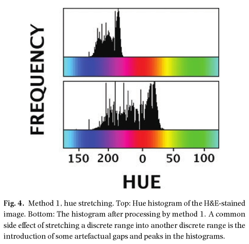
The intent of Method 1 is to create a modified image range that for normal observers that will span a wider range of colours hues and allow the CB observer to detect the wider range of colours. Method 2, re-colours the individual dyes independently and merges them back again into a new single image. Digital images, specific hardware, and computer software was used to prepare the evaluation samples.
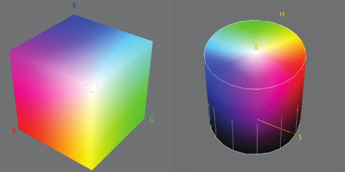
Method 2. The RGB cube colour space (left) and the HSB cylinder colour space. In the RGB cube, a colour is represented by a mixture of red, green and blue components. In the HSB space, a colour is represented by a combination of hue (around the circular base of the cylinder) saturation (less saturated colours are found near the centre) and brightness (increasing from the base to the top).
Results
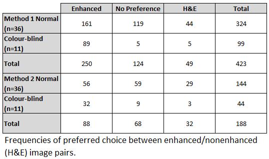
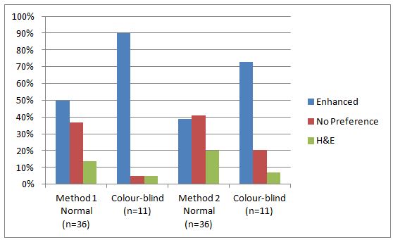
* 36 normal vision and 11 CB individuals volunteered for this study
* 90% CB preferred method 1 over H&E
* 73% CB preferred method 2 over H&E
* 50% Normal Vision preferred method 1 over H&E
* 39% Normal Vision preferred method 2 over H&E
Data suggests method 1 might be preferred however found not statically significant
* 90% CB preferred method 1 over H&E
* 73% CB preferred method 2 over H&E
* 50% Normal Vision preferred method 1 over H&E
* 39% Normal Vision preferred method 2 over H&E
Data suggests method 1 might be preferred however found not statically significant
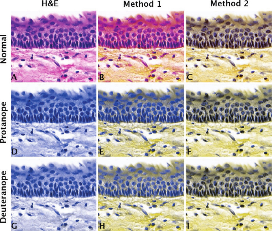
(A) H&E image, (B) enhanced image for red–green CB (method 1), (C) enhanced image for red–green CB (method 2). (D) Protanope simulation of H&E, (E) image enhanced with method 1 as seen by a protanope, (F) image enhanced with method 2 as seen by a protanope-enhanced image D as seen by protanope. (G) Deuteranope simulation of H&E, (H) image enhanced with method 1 as seen by a deuteranope, (I) image enhanced with method 2 as seen by a deuteranope. Note the improvement of the differential rendering of the dyes in images E and F in comparison with D, and in images H and I in comparison with G. Severe protanopes would perceive no differences between A and D, and severe deuteranopes would not perceive differences between A and G.
Conclusion
H&E stain technique is very old and is still the preferred histopathological staining method. Evaluators with CB may not be able to discriminate some important H&E hues. Colour vision deficiency testing is generally not a requirement for laboratory technician careers. This suggests that safe procedures should be put in place to avoid potential errors interfering with diagnostic procedures. The image enhancement methods reported in this research could provide a solution for addressing this problem. Further research using real-time processing of video digital signals to covert images to a live video stream to accommodate an individual’s CBs may be the future of H&E analysis. The normal vision observer would also benefit from the technology with the enhanced images.
